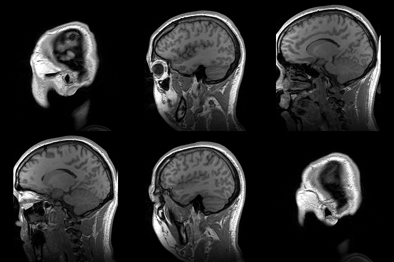
There is huge number of sites which offer free anatomical images in different qualities, but they cannot be an alternative to the Handheld Atlas like Netters etc. The only reason someone may try to find anatomy related images is if its in 3D.
3D is three dimensional. An example of computer generated 3D image is here:
Lets look at few reosurces available on internet for 3D Anatomy:
Visible Human Project:
Website: http://www.nlm.nih.gov/research/visible/visible_human.html
It is developed by National Library of Medicine and National Institute of Health USA.
What does this mean?
It means that they have imaged a whole male and female from top to bottom and are sharing it free with the world. According to wikipedia the male was a murderer who was consented before death for this work but very little is known about the female.
I still do not Understand?
ok than watch this:
How to access it?
Getting your hands on this resource is Free of cost but it requires some effort, you will have to do two things:
More info is here: Getting the Data
Once they approve it you will have a username and password to access over 19 GB of files over there ftp site. There are cross sections 3D images of Brain, Chest (Thorax), Abdomen, Arms (Upper Limb), Pelvis, Genitalia, Legs (Lower Limbs) both of male and female in this dataset provided by NLM and lot more.
Google Body:
Website: http://bodybrowser.googlelabs.com/
There is Google Body as well, which is fast even at 1 mbps and it provides stunning 3D views of human anatomy both male and female.
Wikipedia:
Website: www.en.wikipedia.org
Yes wikipedia also has a huge number of images available and good news is that they can be used in our own works as well, like the example below:
3D is three dimensional. An example of computer generated 3D image is here:
 |
You can see objects behind and in front of each other, this is a 3D image |
Lets look at few reosurces available on internet for 3D Anatomy:
 |
| Visible Human Project |
Website: http://www.nlm.nih.gov/research/visible/visible_human.html
It is developed by National Library of Medicine and National Institute of Health USA.
The Visible Human Project® is an outgrowth of the NLM's 1986 Long-Range Plan. It is the creation of complete, anatomically detailed, three-dimensional representations of the normal male and female human bodies. Acquisition of transverse CT, MR and cryosection images of representative male and female cadavers has been completed. The male was sectioned at one millimeter intervals, the female at one-third of a millimeter intervals.
What does this mean?
It means that they have imaged a whole male and female from top to bottom and are sharing it free with the world. According to wikipedia the male was a murderer who was consented before death for this work but very little is known about the female.
I still do not Understand?
ok than watch this:
How to access it?
Getting your hands on this resource is Free of cost but it requires some effort, you will have to do two things:
- Tell NLM about your intended purpose, they allow commercial use even if the images are used in your own application
- You will have to send them an application in any format PDF, WORD or TXT filled and signed by responsible authority of your instituion.
More info is here: Getting the Data
Once they approve it you will have a username and password to access over 19 GB of files over there ftp site. There are cross sections 3D images of Brain, Chest (Thorax), Abdomen, Arms (Upper Limb), Pelvis, Genitalia, Legs (Lower Limbs) both of male and female in this dataset provided by NLM and lot more.
 |
| Google Body in Chrome Browser |
Google Body:
Website: http://bodybrowser.googlelabs.com/
There is Google Body as well, which is fast even at 1 mbps and it provides stunning 3D views of human anatomy both male and female.
Google Body is a detailed 3D model of the human body. You can peel back anatomical layers, zoom in, and navigate to parts that interest you. Click to identify anatomy, or search for muscles, organs, bones and more.
You can also share the exact scene you are viewing by copying and pasting the corresponding URL.
You will need a web browser that supports WebGL, such as Google Chrome.
Wikipedia:
Website: www.en.wikipedia.org
Yes wikipedia also has a huge number of images available and good news is that they can be used in our own works as well, like the example below:
 |
| Para-sagittal MRI of the head in a patient with benign familial macrocephaly. By Dwayne Reed at en.wikipedia |

Ackland's Video Atlas is best.
ReplyDeletePrimal Pictures is also good but you can't rotate it.
I used Google Body in 2nd yr but it was not quite impressive.
Visible human project is good but I think it is intended to specialists not med students.